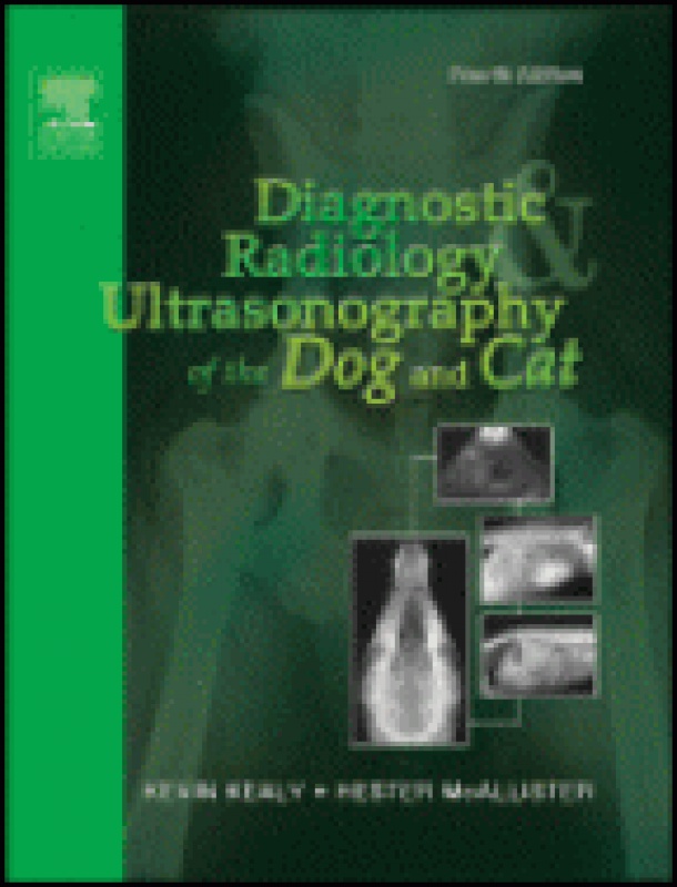
Description
This essential guide to the interpretation of radiographs and
ultrasonograms illustrates normal anatomy and frequently encountered
abnormalities and anomalies. It provides easy access to the
fundamentals of image interpretation with thorough discussions of the
basic principles and techniques for producing high quality radiographs.
Coverage includes the principles of ultrasound and its interaction with
tissue, as well as how to avoid problems that can result from
misinterpretation or overinterpretation of images. It provides a
comprehensive review of canine and feline radiographic and
ultrasonographic anatomy using a systems approach, focusing on normal
features, so that abnormalities can be readily appreciated and
interpreted.
Contents
1. THE RADIOGRAPH
Density And Opacity
Contrast
Radiologic Changes
Standard Views
Contrast Media
Viewing the Radiograph
Ultrasound
2. THE ABDOMEN
The Abdominal Cavity
The Abdominal Wall
The Retroperitoneal Space
The Liver
The Gallbladder
The Spleen
The Pancreas
THE ALIMENTARY TRACT
Esophagus
The Stomach
The Small Intestine
The Large Intestine
The Adrenal Glands
THE URINARY SYSTEM
The Kidneys
The Ureters
The Bladder
The Urethra
THE MALE GENITAL TRACT
The Penis
The Testes
The Prostate Gland
THE FEMALE GENITAL TRACT
The Uterus
The Ovaries
The Vagina
The Mammary Gland
3. THE THORAX
The Thoracic Cavity
The Pharynx, Larynx, and Hyoid Apparatus
The Trachea
The Bronchi
The Lungs
The Diaphragm
The Pleurae
The Mediastinum
The Thoracic Wall
The Spine
The Ribs
The Sternum
The Skin
The Cardiovascular
System
4. BONES AND JOINTS
Bones
Joints
5. THE SKULL AND VERTEBRAL COLUMN
The Skull
The Nasal Chambers
The Paranasal Sinuses
The
Auditory System
The Eye
The Teeth
The Salivary Glands
The Nasolacrimal Ducts
The Brain
The Vertebral Column
The Intervertebral Discs
6. SOFT TISSUES
Calcification (Mineralization)
Arteriovenous Fistula
Fascial Planes
Soft Tissue Pathology
Cervical Soft Tissues
Thyroid Gland
The Parathyroid Glands
Muscles
Lymph Nodes